Title of paper under discussion
Activated brain regions in musicians during an ensemble: a PET study
Authors
Masayuki Satoh, Katsuhiko Takeda, Ken Nagata, Jun Hatazawa, Shigeki Kuzuhara
Journal
Cognitive Brain Research, 12 (2001), pp101–108
Link to paper (free access)
Overview
There are many different ways of listening to music, for instance with attention directed toward the harmony as a whole, or honing in on one particular line within the score. Prof Satoh and colleagues recruited music students from Akita University, Japan to investigate if such different modes of listening involved different patterns of activity in the brain. Whilst playing Bruckner motets through participants’ earphones and directing them either to pay attention to the overall harmony or solely to the alto line within the score, the researchers took images of their listening brains.
When concentrating on the alto line, blood flow increased to four brain regions – superior parietal lobules, precunei, premotor areas and orbitofrontal cortices – areas associated with attention and analysis. Concentrating on the harmony as a whole stimulated increased blood flow to four different regions – temporal poles, anterior cingulate gyrus, occipital cortex and medial cerebellum – areas associated with music perception and recognition.
Method
Nine music students were injected with radioactive tracers and asked to lie down in a PET (positron emission tomography) scanner. Three four-part Bruckner motets, pre-recorded by a pianist, were played through their earphones as their brains were being scanned, and the participants were asked to perform two tasks in turn as they listened: 1) Harmony-listening task – “listen to the harmony as a whole, and if you hear a minor chord make a sign with the index finger of your right hand” and 2) Alto-part-listening task – “listen to and concentrate on the alto line; if you hear the tonic or dominant note in that line then make a sign with the index finger of your right hand“.
Each experiment was ordered thus: Motet one, task 1 then task 2; Motet two, task 1 then task 2; Motet three, task 1 then task 2.
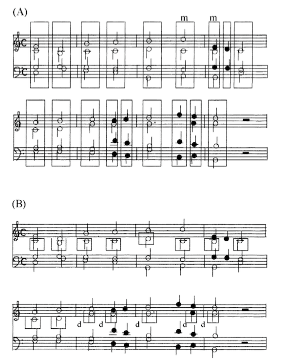
Here’s an example of the tasks illustrated by an excerpt of one of the motets:
Results
Our music students did well in the tasks (recognising minor chords; recognising tonic or dominant in the alto line) responding correctly around 70% of the time.
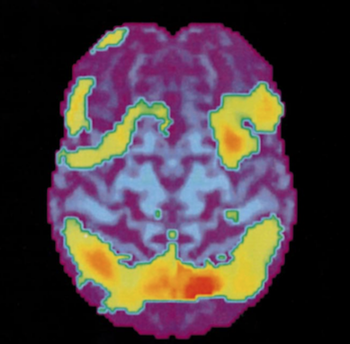
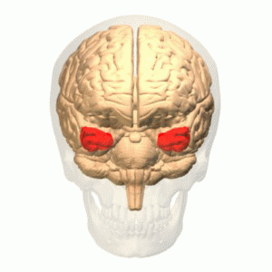
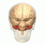
The brain imaging during those tasks is presented in reverse order, task 2 then task 1. The ‘brain activity map’ for task 2 (below) highlights those regions that are significantly more active during alto-part-listening as compared with during harmony-listening:
Looking at these scans (and all will be explained in the ‘discussion’ below), brain anatomists identified that “the alto-part-listening condition produced increases in blood flow in bilateral [both sides of the brain] superior parietal lobules, bilateral precunei, bilateral pre-motor areas and bilateral orbital frontal cortices, compared with the harmony-listening condition“.
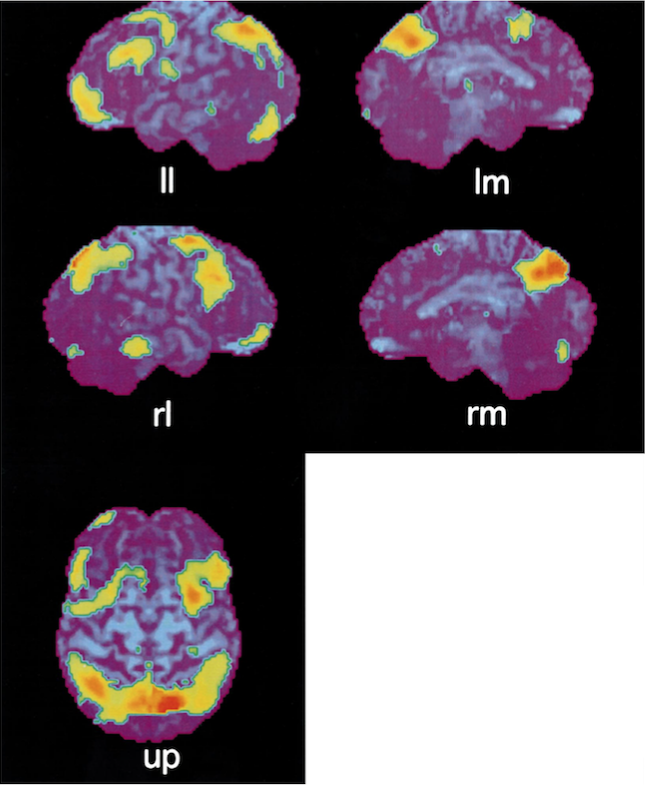
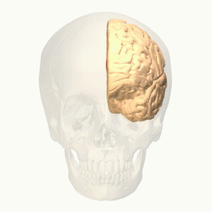
The brain activity map for task 1 (below), in contrast, highlights those regions that are significantly more active during harmony-listening as compared with during alto-part-listening:
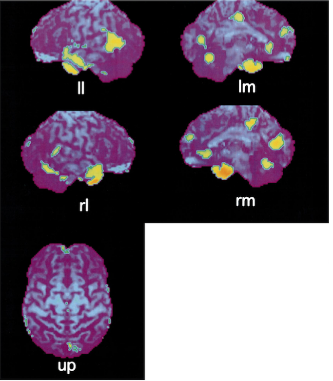
Revealed here is “significantly greater activation” in “bilateral temporal poles, bilateral cingulate gyri, bilateral occipital cortices and medial surface of bilateral cerebellum”.
Discussion
Alto-line listening
Four brain regions ‘lit up’ as the students were concentrating on the alto line; from previous research each area is known to be associated either with ‘paying attention’ or ‘analysing’. These regions are, in turn:
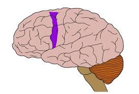
- Superior parietal lobule
According to the authors, previous research suggests that “superior parietal lobules may be activated when the subject selects and pays attention to a part of the target during both visual and auditory processing.” As listeners pay attention to the alto line, ready to spot tonics and dominants, and presumably imagining its progress on a mental stave, both temporal (timing) and spatial attention are important. Temporal attention is thought to be housed mainly in the left parietal lobe, and spatial attention in the right – hence, suggests Satoh, both lobes showing activity as the alto line is attended to.
Another previous paper revealed that the left superior parietal lobe anyhow becomes active when musicians are silently score-reading, apparently due to the mental spatial analysis of notes on a stave. This would tie in with our alto-line-reading students’ activity, paying attention to a line and then analysing its pitch movement.
- Precuneus
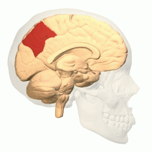
This brain region, on both left and right hemispheres, is also commonly associated with paying attention. In addition to this, the left precuneus is also associated with mental imagery, so perhaps, suggest the authors, its activity in our experiment reflects the listeners’ act of ‘writing the alto line ’ onto a mental score.
- Premotor and orbitofrontal cortex
Again, both these regions have been previously implicated in ‘selective attention’ and ‘attentional effort’. In addition the left premotor area has been shown to be involved in tonal-verbal associations – perhaps our alto-line listeners were mentally naming the notes as they prepared to raise their fingers to identify tonics and dominants.
Harmony-listening
Four brain areas, different from the ones active during alto-line-listening, were activated as the students listened to the motets’ harmonies ‘as a whole’. Such areas have variously, in past research, been associated with music perception and recognition. They were:
- Temporal poles
Patients who lose their temporal poles show “impairment in short term memory for melodies and in discrimination of the metre”, suggesting that these areas are indeed involved in the perception of music.
- Anterior cingulate gyri and occipital cortices
These regions, previous research reveals, are associated with a listener assessing musical familiarity – perhaps, the authors suggest, our students were trying “to judge whether they had ever listened to or were familiar with that musical piece.”
- Cerebella (medial surfaces)
Although the cerebellum is best known for its role in the coordination of movements, it may, according to our authors, “be called into action particularly in anticipation of difficult, new learned tasks in which there is a need for high-quality sensory information.” This would fit the harmony-listening task, which saw each motet being presented for the first time. So, proposes Satoh, “the cerebellum might also have been activated by anticipation of the task that required our subjects to deal with musical pieces that were unknown to them.”

Conclusion
After considering what past research tells us about the functions of brain areas that became active as listeners hone in on an alto line, the authors conclude that “both auditory selective attention and the perception of music via analytic processing allow musicians to listen to a certain vocal part within a harmony”.
Coda
Motet “Os just meditabitur’ WAB 30 – Anton Bruckner
Deutsche Oper Berlin Chorus, conducted by Eugen Jochum
Surprisingly interesting , thanks